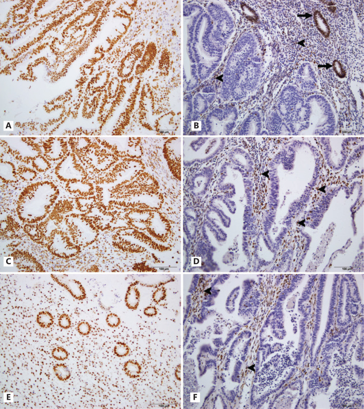
Figure 1.
Expression of MMR protein in EEC. A. MSH2 retained in EEC; B. MSH2 was deficient in EEC; C. MSH6 expressed in EEC; D. Loss of MSH6 in EEC; E. MLH1 retained in endometrial cells; F. MLH1 was lost in EEC. Normal stroma cells (arrowheads) and endometrial cells (arrows) served as internal positive control.

