2.
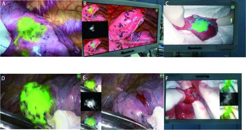
荧光腔镜下肺结节视图。A、D:进胸腔后荧光腔镜下肺结节呈荧光绿染; B、E:荧光腔镜3种模式视图下肺结节呈现图;C、F:肺结节行楔形切除术后,在荧光下呈绿色,切开可明显见肺异常组织
Lung nodules under fluorescent endoscope. A and D: pulmonary nodules showed fluorescent green staining under fluorescent endoscopy after admission into the chest cavity. B and E: pulmonary nodules were shown in the three modes of fluorescent endoscopy. C and F: after wedge resection of pulmonary nodules, the lung tissue where the nodules were located was green under fluorescence, and abnormal lung tissues could be clearly seen after incision.

