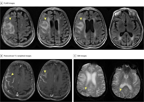
Figure 1. Magnetic Resonance Imaging Findings of Cerebral Amyloid Angiopathy–Related Inflammation.
Magnetic resonance imaging images are shown for a 79-year-old woman who presented with seizure. A, Fluid-attenuated inversion recovery (FLAIR) images show right-sided asymmetric subcortical regions of hyperintensity suggestive of subcortical edema. B, Postcontrast T1-weighted images show right-sided, primarily leptomeningeal contrast enhancement. C, The same right-sided predilection was also noted for foci of cortical superficial siderosis and microbleeds seen on susceptibility-weighted imaging (SWI) images.

