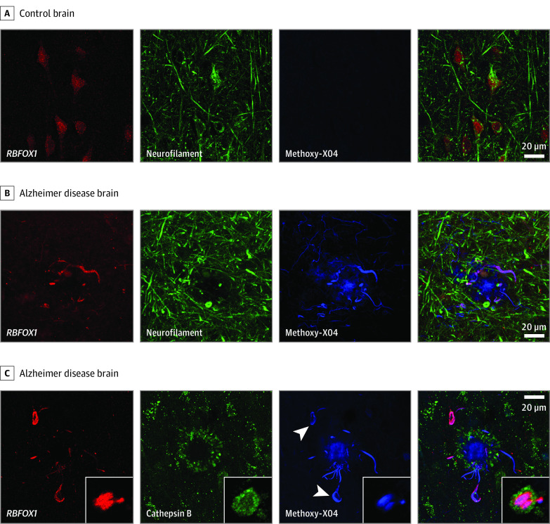
Figure 2. Microscopy of RBFOX1, Neuropil Threads, and Neurofibrillary Tangles .
A, In postmortem control human brain tissue, RBFOX 1 (red) is localized to neurons (neurofilament, green). B, In Alzheimer disease brain, RBFOX1 localizes to neuropil threads around β-amyloid plaques (methoxy-X04, blue). C, In Alzheimer disease brain, RBFOX1 is present in tau tangles (arrowheads) and neuropil threads running through dystrophic neurites (cathepsin B, green) surrounding β-amyloid plaques (methoxy-X04, blue). Insets: cross-section through a dystrophic neurite showing lysosomes (green) surrounding a core of tau (blue) on which RBFOX1 (red) is enriched. Scale bar is 20 μm. Width of inset box is 12 μm.

