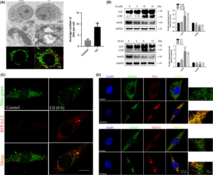
FIGURE 1.

Cd induces mitophagy in PC12 cells. A, TEM analysis of autophagosomes containing mitochondria damaged by Cd (black arrows); average number of AVs (autophagosomes and autolysosomes) quantified per cell; m, mitochondrial; AV, autophagic vacuoles. To examine the connection between mitochondria and autolysosomes, the colocalization of LysoTracker Red with MitoTracker Green in PC12 cells was observed by laser scanning confocal microscopy. Red, LysoTracker Red; green, MitoTracker Green; gold, merge. The gold puncta were considered as autophagosomes containing mitochondria and counted. B, Representative immunoblots and quantification analysis of LC3 and TOM20 in Cd‐treated PC12 cells. GAPDH was used as the internal control. C, Mitochondrial localization of LC3 in PC12 cells increased upon Cd treatment, as indicated by confocal scanning microscopy. Red, RFP‐LC3; green, MitoTracker green (scale bars: 10 μm). D, The LC3 adapters P62 and NDP52 were recruited to mitochondria in PC12 cells upon Cd treatment. The colocalization of P62 with the mitochondrial marker COX IV in PC12 cells was observed by laser scanning confocal microscopy. Red, P62; green, COX IV; gold, merge. The colocalization of NDP52 with COX IV in PC12 cells was observed by laser scanning confocal microscopy. Red, NDP52; green, COX IV; gold, merge. The orange‐yellow puncta were considered as mitochondria containing NDP52 and counted. A minimum of 50 cells were analysed for each experiment. (*P < .05, **P < .01 versus control)
