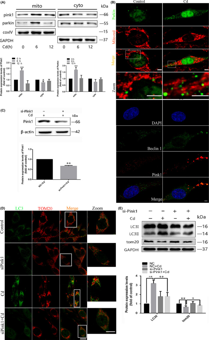
FIGURE 2.

The PINK1/Parkin pathway is involved in Cd‐induced mitophagy in PC12 cells. A, Western blot analysis of PINK1 and Parkin expression in mitochondria and cytoplasm in PC12 cells after Cd exposure. GAPDH and COX IV were used as loading controls. B, Confocal scanning microscopy images of PC12 cells transfected with a rat Parkin‐GFP fusion protein and labelled with MitoTracker Red. Increased mitochondrial localization of Parkin is observed. Colocalization of Beclin1 immunofluorescence and MitoTracker Red fluorescence was also assessed by confocal microscopy analysis. Red, PINK1; green, Beclin1; gold, merge. C, A representative immunoblot of PINK1 protein levels in PC12 cells following PINK1 knock‐down using a commercial si‐PINK1 vector. β‐actin was used as the internal control. D, The colocalization of LC3 dots with TOM20 in PC12 cells following PINK1 knock‐down with si‐PINK1 and Cd treatment indicates mitochondrion containing autophagosome formations. Red, TOM20; green, LC3; gold, merge. The golden puncta were considered as mitochondrion containing autophagosomes and counted. E, LC3 and TOM20 levels in PC12 cells cultured with Cd in the presence or absence of si‐PINK1 were analysed by Western blot. GAPDH was used as the loading control. (scale bars: 1 μm, *P < .05, **P < .01)
