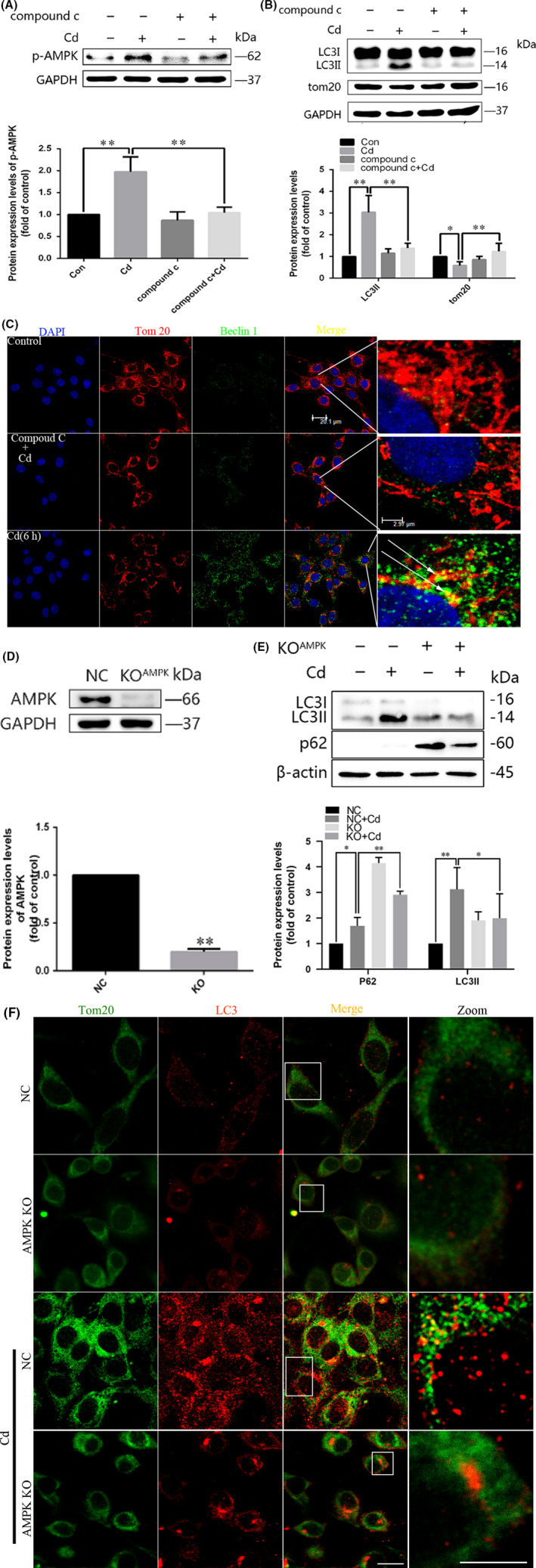
FIGURE 3.

Pharmacological inhibition of AMPK activation decreases the mitochondrial localization of Beclin1 upon Cd treatment. A and B, Representative immunoblots and quantification analysis of phosphorylated AMPK (p‐AMPK), LC3, and TOM20 in Cd‐treated PC12 cells in the presence or absence of Compound C. C, After treatment with 10 μmol/L Cd for 6 h with or without Compound C, the cells were stained for Beclin1 and TOM20 and analysed by confocal microscopy. Red, TOM20; green, Beclin1. D, The AMPK gene was knocked out in PC12 cells using the CRISPR‐Cas9 system. Control cells and AMPK knockout cells were treated with Cd for 6 h, and AMPK levels were measured by Western blot. E, Effects of treatment with 10 μM Cd on LC3‐II and p62 protein levels, as measured by Western blot in AMPK knockout cells. F, The colocalization of LC3 dots with TOM20 was analysed in AMPK knockout cells following Cd treatment to visualize the mitochondrion containing autophagosome formations. Red, TOM20; green, LC3; gold, merge. (scale bars: 10 μm, *P < .05, **P < .01)
