
Figure 4.
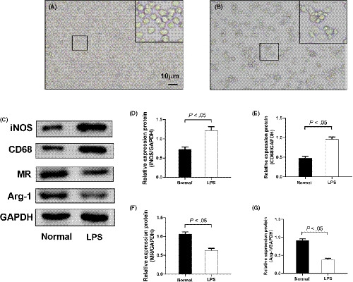
PMA stimulated differentiation of THP‐1 into macrophages, and LPS induced further differentiation of M1 macrophages. A, Morphology of THP‐1 cells. B, Morphology of PMA‐stimulated THP‐1 cells. C‐G, Protein levels of iNOS, CD68, MR and Arg‐1 were detected by Western blotting. Data are shown as mean ± SD. n = 3