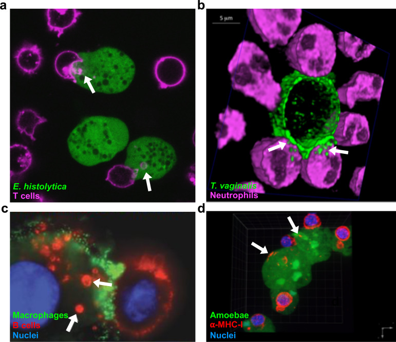
FIG 2.
Examples of trogocytosis within and between species. (a) E. histolytica kills human cells through trogocytosis. E. histolytica is stained with cell tracker green, and human Jurkat T cell membranes are stained with DiD (pink). Arrows, ingested bites. (b) Neutrophils kill T. vaginalis through trogocytosis. T. vaginalis membranes are stained with streptavidin-488 (green), and neutrophils are stained with cell tracker deep red (pink). Arrows, ingested bites. (c) Macrophages can perform trogocytosis to kill antibody-opsonized cells. Macrophages are stained with anti-CD45 (green), Raji B cells are opsonized with trastuzumab (red), and nuclei are stained with Hoechst stain (blue). Arrows, ingested bites. (d) E. histolytica acquires and displays human cell membrane proteins through trogocytosis. E. histolytica is stained with cell tracker green, human anti-MHC-I is shown in red, and nuclei are stained with 4′,6′-diamidino-2-phenylindole (DAPI) (blue). Arrows, acquired MHC-I. (Reprinted from references 20, 41, and 96 with permission.)

