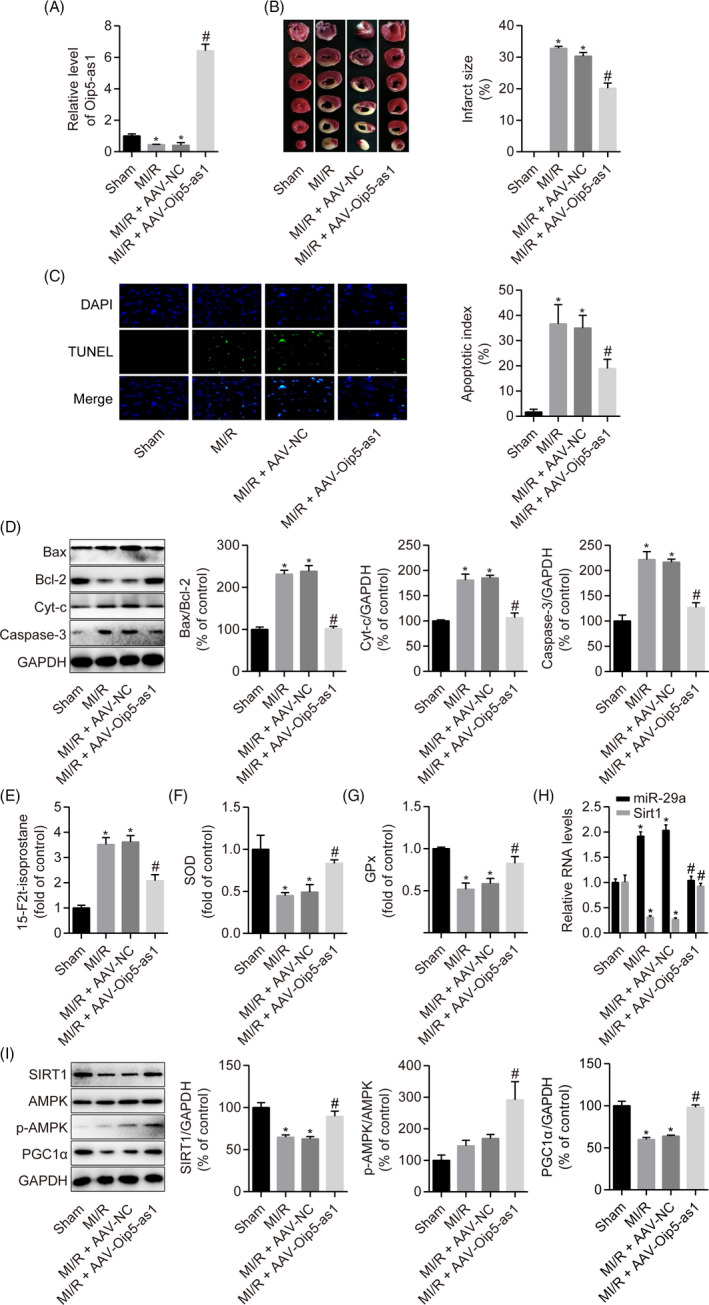
FIGURE 9.

Oip5‐as1 upregulation ameliorates MI/R injury in rats. A, RT‐qPCR analysis of the relative Oip5‐as1 expression levels in rat hearts. B, Representative images of heart sections stained with TTC and the quantitative analysis of myocardial infarct size. C, Representative pictures of cardiomyocyte apoptosis determined by the TUNEL assay and corresponding quantitative analysis. D, Western blot and densitometric analysis of the expression of apoptosis marker proteins (Bax, Bcl‐2, Cyt‐c and Caspase‐3) in rat hearts. E‐G, Measurement of oxidative stress markers (15‐F2t‐isoprostane, SOD and GPx) using the respective assay kits in rat hearts. H, RT‐qPCR analysis of the relative expression levels of miR‐29a and Sirt1 in rat hearts. I, Western blot and densitometric analysis of SIRT1, p‐AMPK and PGC1α levels in heart samples from rats. Data are expressed as mean ± SD (n = 6). *P < .05 vs the sham group. # P < .05 vs the MI/R + AAV‐NC group
