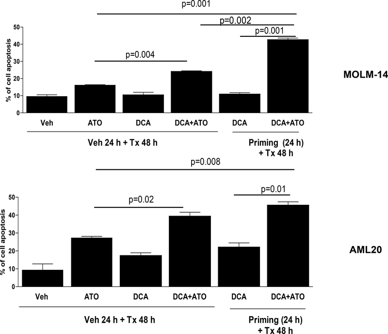
Figure 3. Comparison of apoptosis in AML cells with different treatment strategies.
Treatment of MOLM-14 and AML20 cells with combination of DCA at their corresponding IC30s and ATO at their corresponding IC50s with priming strategy as described in Methods and Materials significantly (p<0.05) increased apoptosis compared to ATO alone or DCA alone or their combination without priming.

