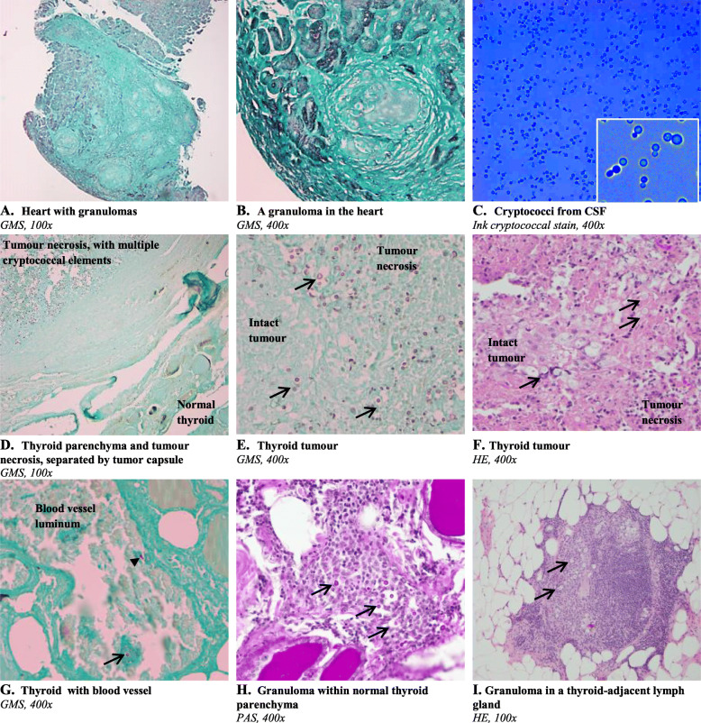
Fig. 2.
Histological sections and cerebrospinal fluid microscopy. Arrows indicate characteristic cryptococcal elements with the capsule appearing as an unstained clear halo. Endomyocardium with granulomas but with no evidence of cryptococci a and b, CSF finding of cryptococci c, thyroid tumour necrotic area with cryptococci but with no signs of cryptococci in the adjacent normal thyroid glandular tissue d, thyroid tumour necrosis and focal intact tumour tissue e, thyroid tumour with cryptococci and inflammatory infiltrate f, thyroid parenchyma without cryptococci except for within a blood vessel, note budding indicated by the arrowhead g, cryptococcal elements in epithelioid cell granulomas in the thyroid parenchyma h, cryptococcal elements in granulomas in a thyroid-adjacent lymph gland i. Staining types are cryptococcal ink, hematoxylin and eosin (HE), the fungal-specific Grocott’s methenamine silver (GMS) stain and Periodic acid–Schiff (PAS)

