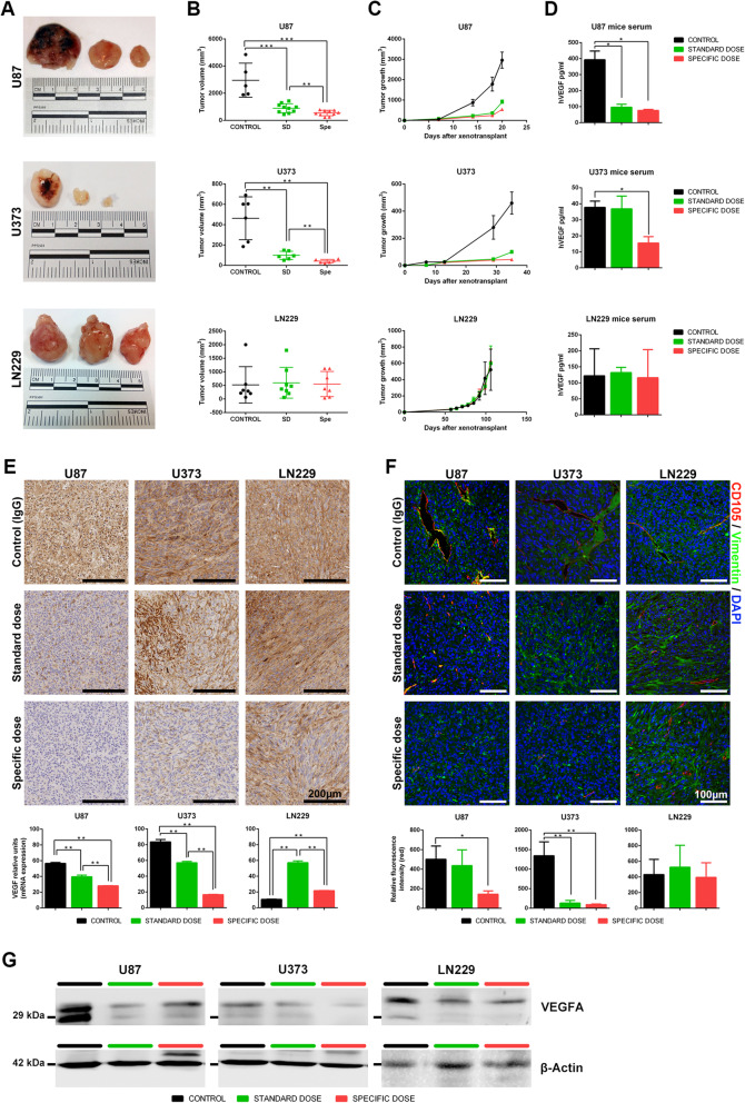
Fig. 5.
In vivo tumor growth inhibition by a specific dose of bevacizumab in high VEGFA secreted cell lines. a 1.5 × 10⁶ U87, U373, and LN229 cells were implanted into the flanks of nude mice. Mice were treated with IgG (n = 10), SD (n = 5), and a specific dose of bevacizumab (n = 5) twice a week. Representative macroscopic tumor images of control (left), SD, and Spe (right) are shown. b Tumor volume (mm3) at the endpoint. c Tumor growth curves. d Human VEGFA measure (pg/ml) from mouse serum. e Immunohistochemistry images using anti-VEGFA antibody, scale bar 200 μm, and real-time PCR analysis assessing VEGFA expression in tumoral tissue. f Immunofluorescence microscopy using anti-human vimentin (green) and mouse CD105 (red). The nuclei were stained with DAPI (blue). Scale bar, 100 μm. Relative fluorescence intensity was calculated for CD105. g Western blots demonstrating that Spe of bevacizumab inhibits VEGFA in U87 and U373 cell lines. Results are shown as mean ± S.D. *P < 0.05, **P < 0.01, ***P < 0.001

