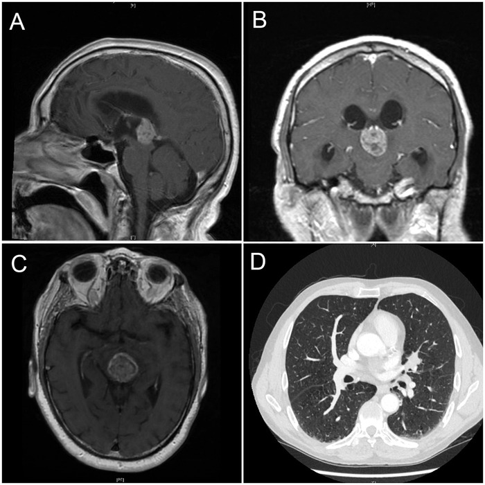
Figure 1.

(A) Sagittal sections of magnetic resonance imaging (MRI) of the brain showing the pineal gland mass. (B) Coronal and (C) transverse sections of brain MRI showing the same pineal tumor. (D) Computed tomography of the chest with intravenous contrast showing a lung nodule over the left hilar region in the lower right corner.
