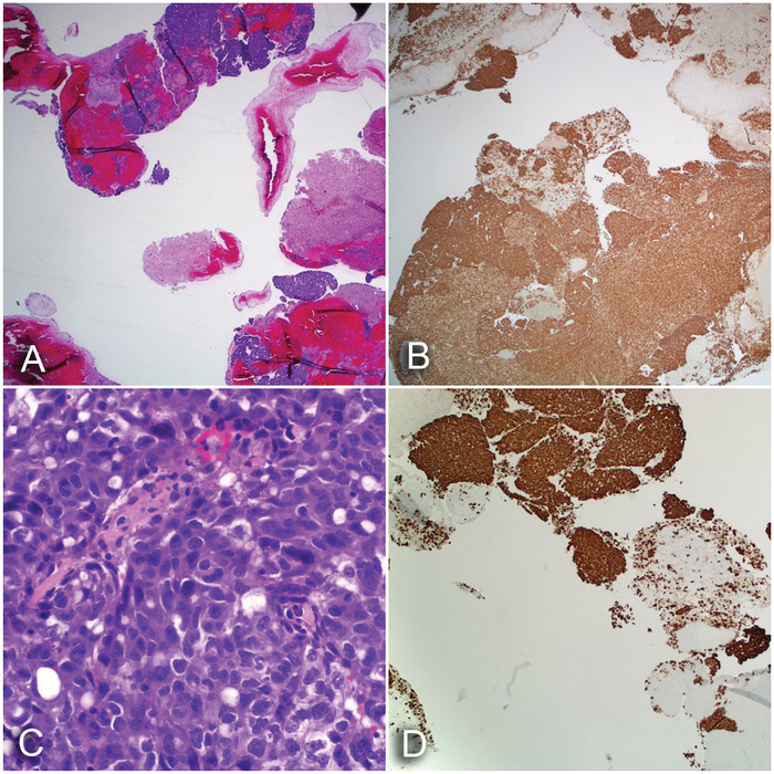
Figure 2.

(A) Epithelioid neoplasm (hematoxylin and eosin [H&E] stain, ×20). (B) Strong and diffuse staining for pankeratin (H&E stain, ×20). (C) Epithelioid neoplasm demonstrating marked nuclear pleomorphism with eosinophilic cytoplasm (H&E stain, ×400). (D) Strong and diffuse staining of the neoplasm for thyroid transcription factor-1, supporting the diagnosis of adenocarcinoma of pulmonary origin (H&E stain, ×40).
