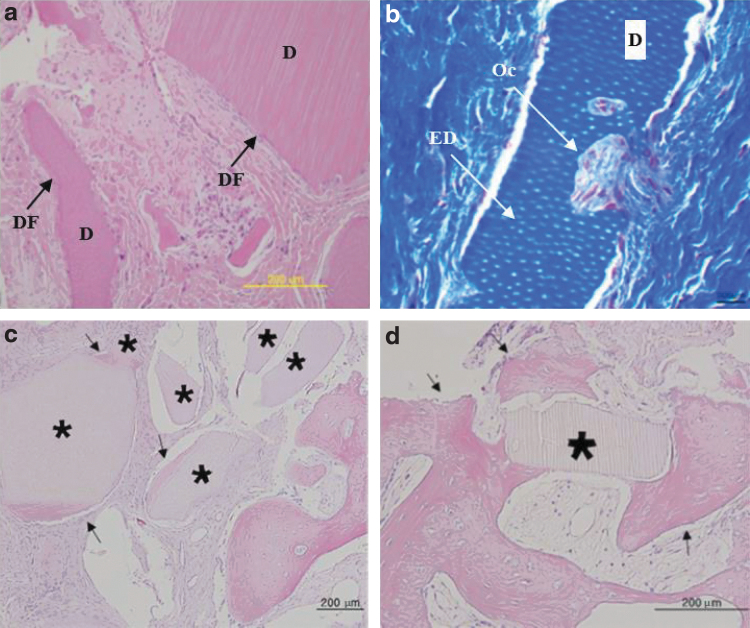
FIG. 4.
Histological features of human DDM and DDM/rhBMP-2 implanted for socket preservation. (a) Biopsy specimens of human DDM alone in mandibles at 3.5 months after the socket preservation procedure (hematoxylin–eosin staining, scale bar = 200 μm) under gingival tissue.29 DDM was surrounded by dense fibrous connective tissues (DF), which were in close contact with the particle, leaving no gaps. The FC seemed to consist of inactive flat fibroblast-like cells. Dentin matrix resorption was not observed. (b) Biopsy specimens of human DDM/rhBMP-2 (0.2 mg/mL; Cowell BMP, Busan, Korea) in the maxilla at 6 months after the socket preservation procedure (hematoxylin–eosin staining, scale bar = 200 μm) under gingival tissue.29 Enlarged dentinal tubules (ED) were observed. Multiple activated cell layers encapsulated the dentin matrix surface. The resorption cavity was infiltrated with osteoclast-like cells (Ocs) that may facilitate the release of ED-BMPs present as matrix- and hydroxyapatite-binding proteins. (c) Trephine biopsy specimens of human DDM alone in the mandible at 3 months after the socket preservation procedure (hematoxylin–eosin staining, scale bar = 200 μm).31 Bone formation (arrows) around the DDM particles (*) surrounded by dense fibrous connective tissue. (d) Trephine biopsy specimens of human DDM DDM/rhBMP-2 (0.2 mg/mL; Cowell BMP, Busan, Korea) in the mandible at 3 months after the socket preservation procedure (hematoxylin–eosin stain, scale bar = 200 μm).31 Active bone formation (arrows) was observed with embedding of osteocytes and lining of osteoblasts around the DDM particles (*), which were surrounded by highly vascularized alveolar tissue. DF, dense fibrous connective tissues.

