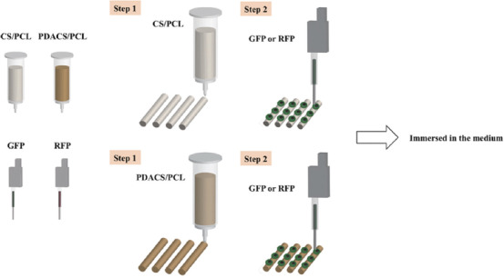
Figure 1.

Schematic diagram of the bioprinting process. First, a framework was fabricated with calcium silicate/polycaprolactone (CS/PCL) and polydopamine CS/PCL composite to support scaffold stability. Second, the cells (or fluorescein isothiocyanate) were printed on the scaffold surface by the piezoelectric needle.
