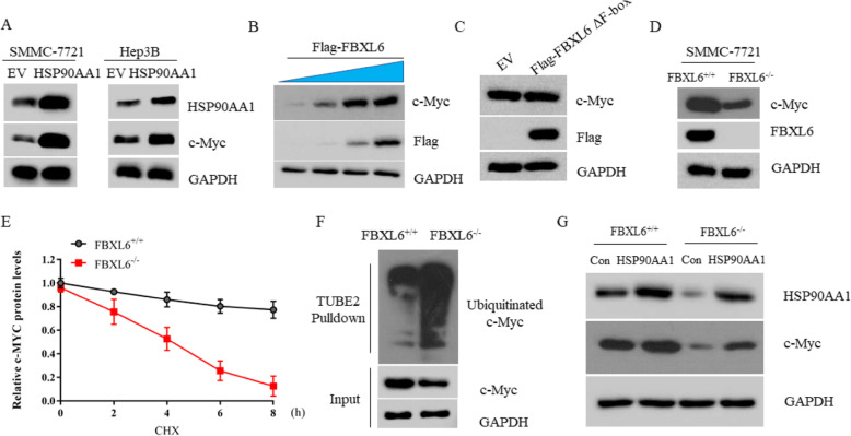
Fig. 5.
FBXL6 stabilizes c-MYC via HSP90AA1. a Western blot analysis of the WCL derived from SMMC-7721 cells or Hep3B cells transfected with EV or HSP90AA1 plasmids. b Western blot analysis of the WCL derived from Hep3B transfected with increased dose of Flag-FBXL6. c Western blot analysis of the WCL derived from SMMC-7721 cells transfected with EV or Flag-FBXL6ΔF-box plasmids. d The protein levels of c-Myc from FBXL6+/+and FBXL6−/− SMMC-7721 cells were detected by immunoblotting. e FBXL6+/+and FBXL6−/− SMMC-7721 cells were treated with 20 μM CHX for the indicated time. The whole cell lysate was immunoblotted with anti-c-Myc antibody. The quantification plot was based on scanning densitometry analysis using the Image J software. Relative protein levels were normalized to FBXL6−/− control group. f The WCL from FBXL6+/+and FBXL6−/− SMMC-7721 cells were immunoprecipitated by Tandem Ubiquitin Binding Entity 2 (TUBE2) resin for ubiquitinated proteins enrichment and immunoblotted as indicated. g FBXL6+/+ and FBXL6−/− SMMC-7721 cells transfected with vector control or HSP90AA1 plasmids were subjected to western blot assay with indicated antibodies

