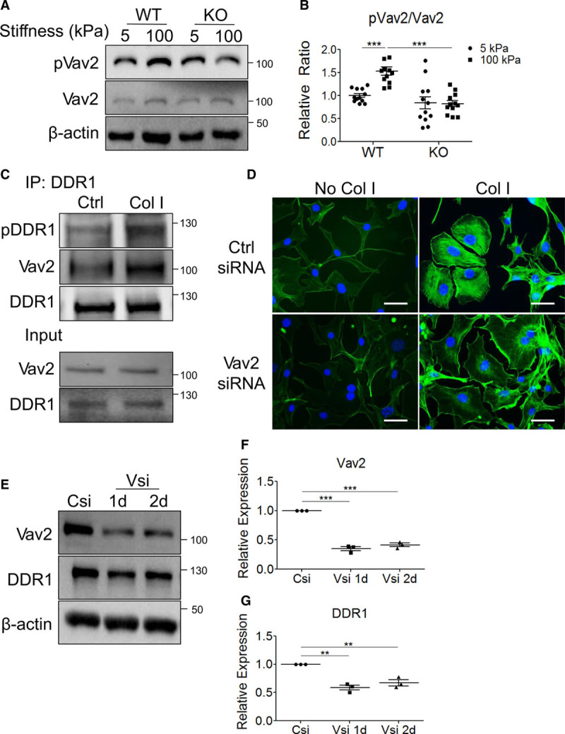
Figure 4.

Vav2 promotes Col-I (collagen-I)–dependent stress fiber formation. A, Wild-type (WT) or knockout (KO) vascular smooth muscle cells (VSMCs) were seeded on 5 or 100 kPa substrates and lysed after 3 h before immunoblotting (n=12). B, pVav2/Vav2 (phosphorylated Vav2 to total Vav2) ratios were quantified by densitometry and expressed relative to 5 kPa WT. C, DDR1 (discoidin domain receptor-1) was immunoprecipitated (IP) using WT VSMCs with or without Col-I stimulation (n=3). D, WT VSMCs transfected with control (Ctrl) siRNA (small interfering RNA; Csi) or Vav2 siRNA (Vsi) were serum-stimulated for 3 h on uncoated coverslips or Col-I–coated coverslips, then stained with phalloidin (n=3). Phalloidin staining was imaged at 400x magnification. Scale bar represents 50 μm. E, WT VSMCs were transfected with Csi or Vsi and lysed after 1 or 2 d (n=3). Quantification of immunoblots in (E) is shown for (F) Vav2 and (G) DDR1, with values expressed relative to Csi. B, F, and G, **P<0.01, ***P<0.001, bars represent means±SEM, and statistics were done by (F and G) 1-way or (B) 2-way ANOVA with Bonferroni post hoc tests.
