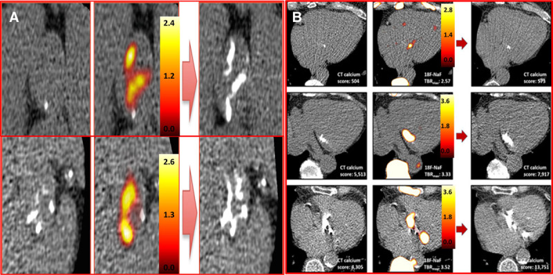
Figure 2.

18F-sodium fluoride (18F-NaF) uptake predicts progression of aortic valve calcification and mitral annular calcification (MAC). A, Baseline calcium score (left) of 2 patients with aortic sclerosis (top) and moderate aortic stenosis (bottom). Fused 18F-NaF positron emission tomography (PET)–computed tomography (CT) scans (middle) show fluoride uptake in red and yellow. Follow-up CT at 2 y (right) indicated new areas of macroscopic calcium on the repeat CT in a similar distribution to the PET activity (reprinted from Jenkins et al47 with permission. Copyright ©2015, the Journal of the American College of Cardiology). B, Baseline calcium score (left) of patients with mild, moderate, and severe MAC. Fused 18F-NAF PET-CT scans (middle) show fluoride uptake in red and yellow. Similar to the aortic valve the baseline PET predicts where new macroscopic calcium in the mitral valve will develop on follow-up CT at 2 y (right) (reprinted from Massera et al.44 Copyright ©2019, the Authors).
