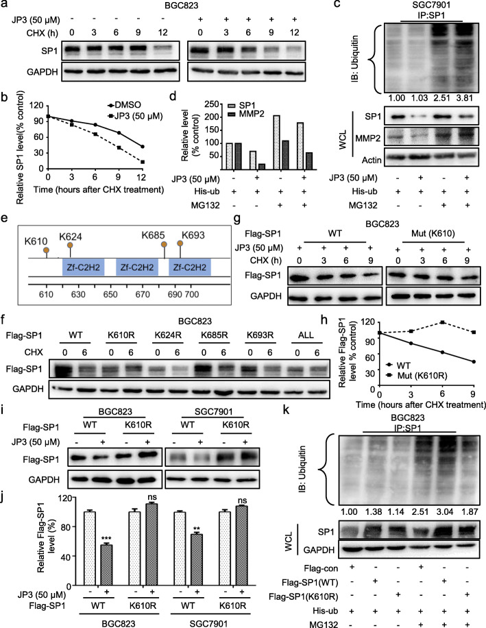
Fig. 3.
JP3 triggers ubiquitination modification of SP1 at K610 in GC cells. a BGC823 cells were treated with JP3 (0 or 50 μM), and then with CHX and harvested at the indicated time points for Western blotting. b The relative intensities of the SP1 protein bands were analyzed by densitometry after normalization to GAPDH. c Ubiquitination of SP1 was induced by JP3. His-ub was transfected into SGC7901 cells for 48 h and with JP3 (0 or 50 μM) for another 24 h, followed by pre-treatment with or without MG132 (10 μM) for 6 h. d The intensities of the SP1 and MMP2 protein bands in SGC7901 cells were analyzed by densitometry after normalization to Actin. e Data from the PhosphoSitePlus (https://www.phosphosite.org) showed the potential sites required for ubiquitination of SP1. f BGC823 cells were transfected with Flag-SP1 (WT) or mutants, followed by exposure to CHX (100 μg/ml) for 6 h. The indicated proteins were detected by Western blotting. g-h BGC823 cells were transfected with Flag-SP1 (WT) or Flag-SP1 (K610R) for 48 h and then JP3 (50 μM) for 24 h, followed by exposure to 100 μg/ml of CHX for 0, 3, 6, 9 h; the protein level of Flag-SP1 was determined by Western blotting, and the intensity of the SP1 protein bands were analyzed (h). i, j BGC823 (left) and SGC7901 (right) cells were transfected with Flag-SP1 (WT) or Flag-SP1 (K610R) for 48 h, followed by treatment with JP3 (50 μM) for 24 h, and j the intensity of the SP1 protein bands were analyzed. The data are presented as the means ± SEM, ns: no significance, **P < 0.01, ***P < 0.001. k BGC823 cells were co-transfected with His-Ub, Flag-SP1 (WT) or Flag-JWA (K610R) for 48 h, followed by pre-treatment with MG132 (10 μm) for 6 h. Ubiquitinated SP1 was determined by IP with anti-SP1. The indicated protein levels were determined by Western blotting

