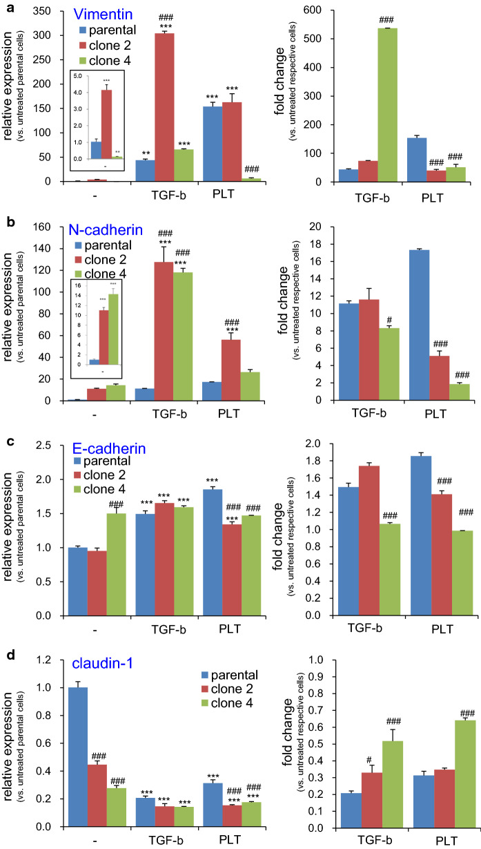
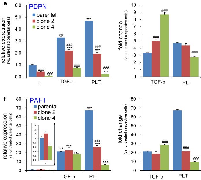
Fig. 6.
Effect of PDPN knockout on PLT- or TGF-β-induced expression of EMT-related genes in TE11A cells. Parental, clone 2, or clone 4 TE11A cells at confluence in 24-well plates were treated with TGF-β (20 ng/mL) or platelets (~ 2 × 10⁷/mL) plus EGTA (2 mM) for 18 h. The RNA was then extracted, and the expression of the EMT-related genes, vimentin (a), N-cadherin (b), E-cadherin (c), claudin-1 (d), PDPN (e), and PAI-1 (f), was analyzed by real-time PCR. In each left side graph, relative level of expression is shown with respect to non-treated parental TE11A clone, whereas in the right side graph, fold induction is expressed with respect to the basal level of each clonal cells. The inset graph in (a, b, f) shows the magnified bars for the basal level of expression of each cell clone. Values represent the mean ± SEM of quadruplicate culture wells from one representative experiment. *P < 0.001 and ***P < 0.001 vs. non-treated parental cells by one-way ANOVA and Tukey’s test, ###P < 0.001 vs. parental cells of respective treatment group by two-way ANOVA and Bonferroni’s test


