Fig. 3.

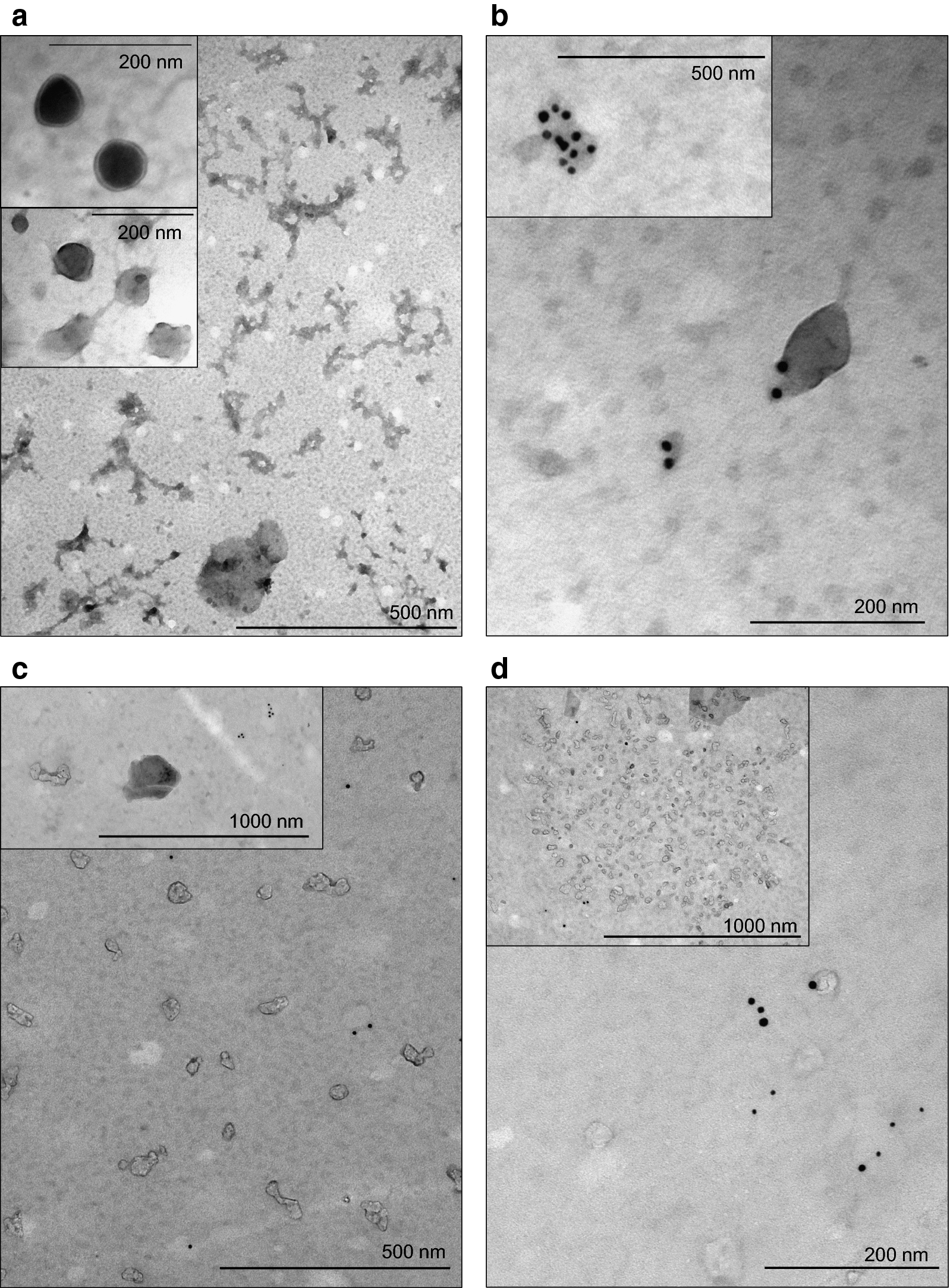
Atypical extracellular vesicles in cerebrospinal fluid of patients with different central nervous system (CNS) status. a Transmission electron microscopy (TEM) images of a cerebrospinal fluid (CSF) sample from a CNS+ patient with precursor B-cell acute lymphoblastic leukemia (pB-ALL) depict several small extracellular vesicles (EVs). Inserted panels show the absence of CD63/CD81 immunogold labelling which is a key feature of these EVs. b TEM images taken from the CSF sample of a patient with CNS‒ pB-ALL. Appearance of gold particles refer to CD63 (10 nm) and CD81 (5 nm) positivity of the EVs. c Small EVs in a patient with relapsed CNS+ T-cell ALL show mainly the absence of CD63/CD81 immunogold labelling. d CD63+/CD81+ small EVs in CSF from a patient with T-cell ALL without CNS involvement. Conversely, inserted slide shows a conglomerate of EVs without typical markers of exosomes originating from multivesicular body
