Fig. 3.
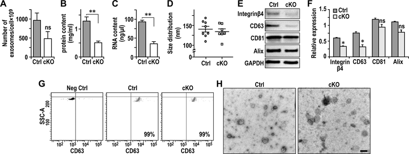
FAK deletion in CAFs results in defective exosomes. A-C. Quantification of (A) numbers of exosome generated per million of cells (volume of preparation), (B) protein content (mg/ml) per volume of preparation and (C) RNA content per volume of preparation of exosomes from purified primary lung fibroblasts. D. Quantification of exosome size distribution by nanosight. E. Immuno-blots showing levels of exosome markers Integrinβ4, CD63, CD81 and Alix using purified exosomes from primary lung fibroblasts of Ctrl or cKO mice. F. Quantification of respective protein levels from purified exosomes normalized against GAPDH as described in E. G. CD63 FACS analysis of purified exosomes. Exosomes without CD63 primary antibody incubation were utilized as negative control. H. Representatives images of electron microscopic analysis of exosomes derived from the primary lung fibroblasts. Scale bar, 100nm. Error bars indicate mean ±SEM. **P<0.01.

