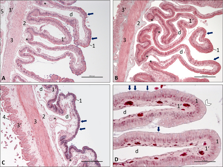
Fig 6. Photomicrographs of the wall of the small intestine, Inland Bearded Dragon (Pogona vitticeps), transverse sections.
(A) duodenum, haematoxylin and eosin staining, bar 250 μm; (B) jejunum, Masson-Goldner staining, bar 250 μm; (C) ileum, haematoxylin and eosin staining, bar 250 μm; (D) duodenum, Masson-Goldner staining, bar 62.5 μm. Mucous tunic consisting of epithelium (1) interspersed with goblet cells (arrows) and lamina propria (1‘) with large central vessel (d); submucosal layer (2); muscular tunic consisting of an inner circular stratum (3) and an outer longitudinal stratum (3’); serosal tunic (4); attachment of mesentery (5). D: The epithelium (1) shows a brush border (white arrowhead). Smooth muscle cells (*) are located near the wall of the central vessel (d) in the lamina propria (1’).

