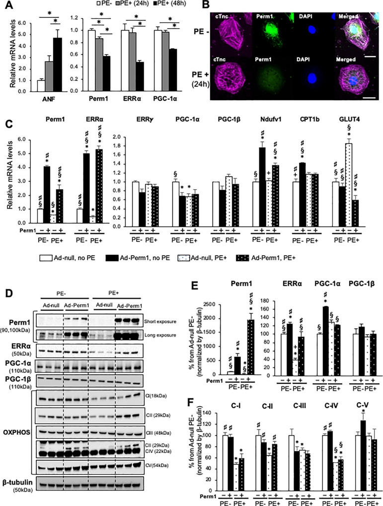
Fig 5. Perm1 is downregulated in cardiomyocytes under cellular hypertrophic stress.
(A) RT-PCR showing Perm1 downregulation in NRVMs treated with phenylephrine (PE) (n = 3/group *:p<0.05 by one-way ANOVA). (B) Immunostaining of NRVMs showing the localization of Perm1 in the nucleus and the peripheral nuclear region (Top). After incubation with PE for 24hr, Perm1 was less localized in the nucleus and showed a diffuse expression pattern. Cardiac muscle-specific anti-troponinC (cTnc) was used to distinguish cardiomyocytes from fibroblasts. Scale bar: 20 μm. (n = 3/group). (C-F) RT-PCR (Panel C) and Western blot analysis (Panels D-F) showing that Perm1 overexpression prior to 48h incubation with PE (group Perm1+/ PE+) either completely or partially rescues downregulation of some metabolic genes during PE-induced hypertrophic stress. The control (group Perm1-/PE-) was obtained by treating NRVMs with adenovirus-null for 48h without incubating with PE. The data for group Perm1-/PE- and group Perm1+/PE- in Panel C is the same as shown in Fig 4A. Note that the expression levels of the Complex II and III subunits were quantified using individual antibodies againt SDHB and UQCRC2 in separate blots due to being adjacent to the band from the relatively highly abundant Complex IV subunit. Statistics were performed using one-way ANOVA. *: p<0.05 compared with Perm1-/PE- (control); §: p<0.05 compared with Perm1+/PE+; #: p<0.05 compared with Perm1-/PE-. Error bars are ±SEM.

