Figure 1.

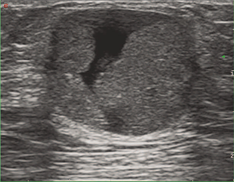
The long-axis sonogram of the left breast showed a regular shaped, well-defined complex mass with the cystic and solid hypoechoic mass in the retro areolar region.

The long-axis sonogram of the left breast showed a regular shaped, well-defined complex mass with the cystic and solid hypoechoic mass in the retro areolar region.