Figure 3.
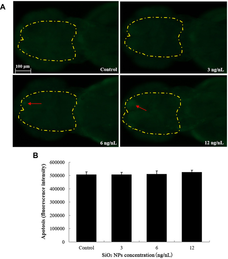
The apoptosis of brain cells induced by SiO2 NPs exposure was determined using acridine orange staining in embryos of zebrafish after a 24 h exposure. (A) The images of apoptosis of brain cells were detected by fluorescence microscope. (B) The relative fluorescence of cellular apoptosis of brain was detected. Data are expressed as means S.D. from three independent experiments.

