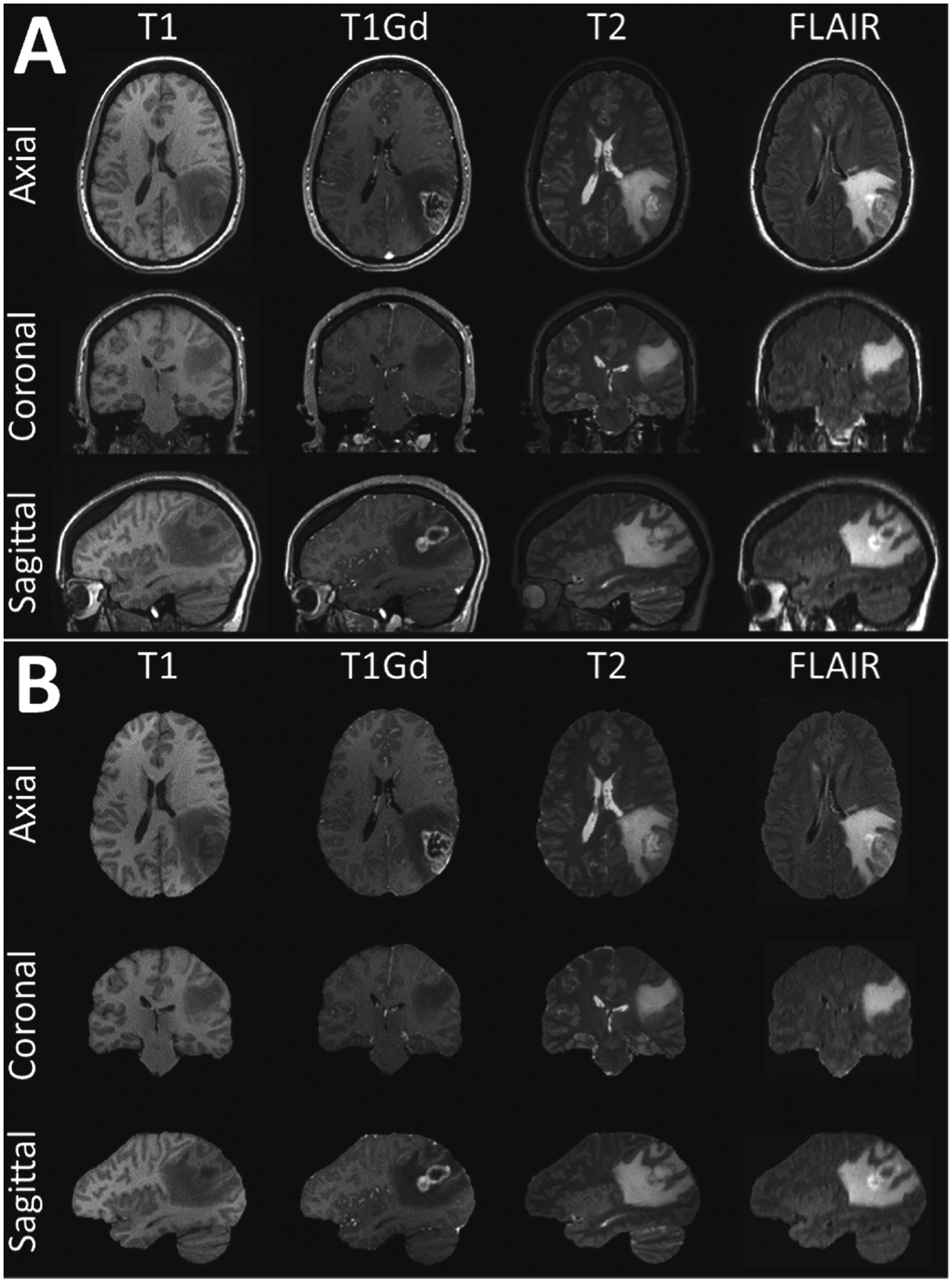
Fig. 1.

Example mpMRI brain tumor scans from a single subject. The original scans including the non-brain-tissues are illustrated in A, whereas the same scans after applying the manually-inspected and verified gold-standard brain tissue segmentations are illustrated in B.
