. 2020 Jun 23;9:e56185. doi: 10.7554/eLife.56185

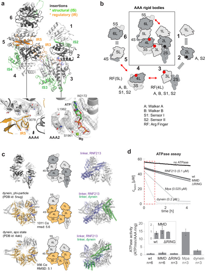
Figure 2. AAA core adopts a dynein-like fold.

(a) Organization of the 6 AAA domains, shown in alternating gray tones, highlighting insertions with structural (IS, green) and regulatory (IR, orange) roles. Orientation is similar as in Figure 1c. The insets below illustrate the EM density of the IR5 motif blocking via Tyr3078 nucleotide binding to AAA4 and the ATP molecule tightly bound at AAA2 (also shown in Video 1). (b) Schematic cartoon of the ATPase core emphasizing the AAA ‘rigid bodies’ formed between the L domain of one and the S domain of the previous unit (Wang et al., 2001). The catalytically competent ATPase sites (red dots), the ATP-bound AAA2 (black dot), and the respective functional motifs for ATP binding and hydrolysis are indicated. (c) Structural superposition of RNF213 (top row) with two dynein states (Phi particle, middle; apo dynein, bottom). The left panel illustrates the aligned dynein domains (orange), the middle shows the corresponding cartoon of the matching RNF213 AAA portions, and the right panel highlights the upstream linker of RNF213 (lilac) superimposed with phi-particle and apo dynein structures (AAA core, grey; linker, green). (d) ATPase assay comparing RNF213 variants with dynein and a processive ATPase, the unfoldase Mpa. The top panel illustrates representative NADH decay curves, with used protein concentrations being indicated. The region for calculating reaction rates is highlighted in red. The lower panel quantifies the respective ATPase rates calculated per AAA ring. Rates for tested RNF213 variants are also shown enlarged. Error bars indicate standard deviation.

Figure 2.

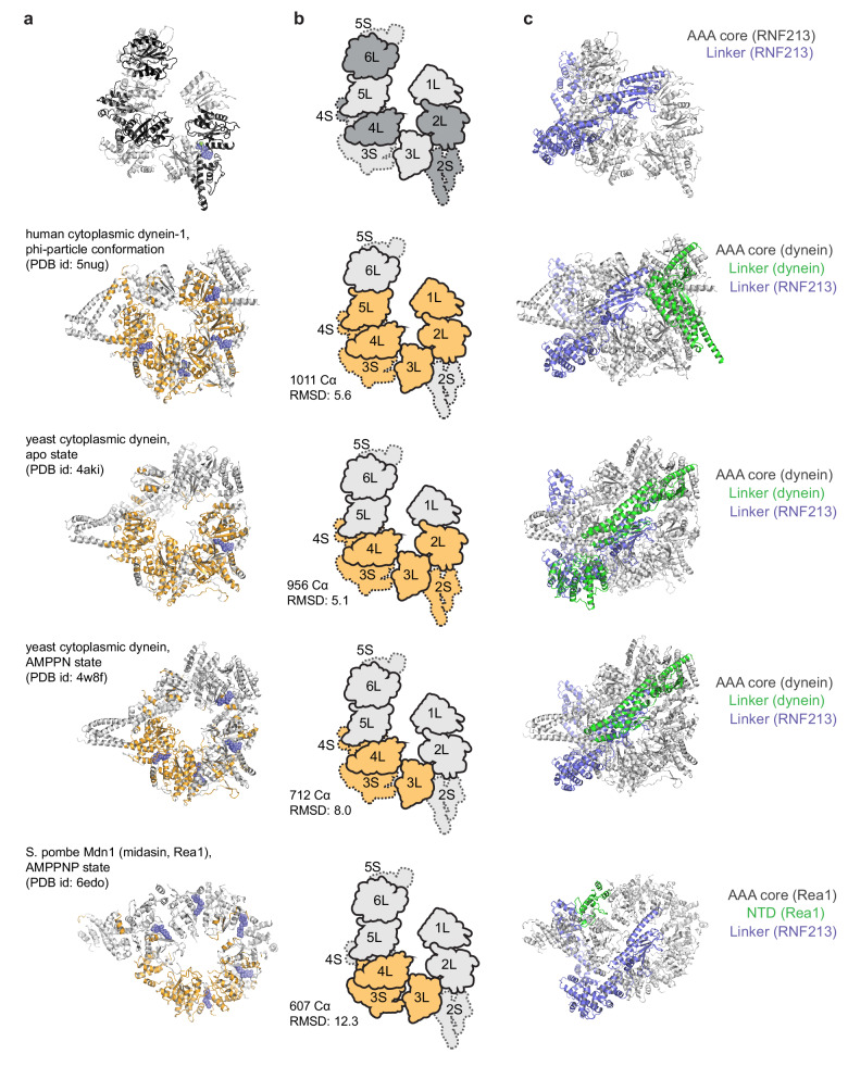
Figure 2—figure supplement 1. Structural comparison of RNF213 with dynein-like proteins, containing 6 AAA units in a single polypeptide.

Figure 2—figure supplement 1.

(a) AAA cores of dynein Phi particle, dynein apo state, dynein AMPPNP-bound form, and Rea1, highlighting those residues in orange that could be aligned to RNF213 (top). The rms deviations and number of aligned Cα atoms are indicated. (b) Schematic cartoon of the RNF213 AAA core, emphasizing the aligned AAA units. Accordingly, RNF213 and dynein show the greatest similarity in the AAA2/3/4 part. (c) AAA cores of RNF213, dynein and Rea1 as in panel a) are shown together with their N-terminal linker (green), superimposed with the RNF213 counterpart (lilac). The pictures highlight the similar linker architecture in RNF213 and dynein.
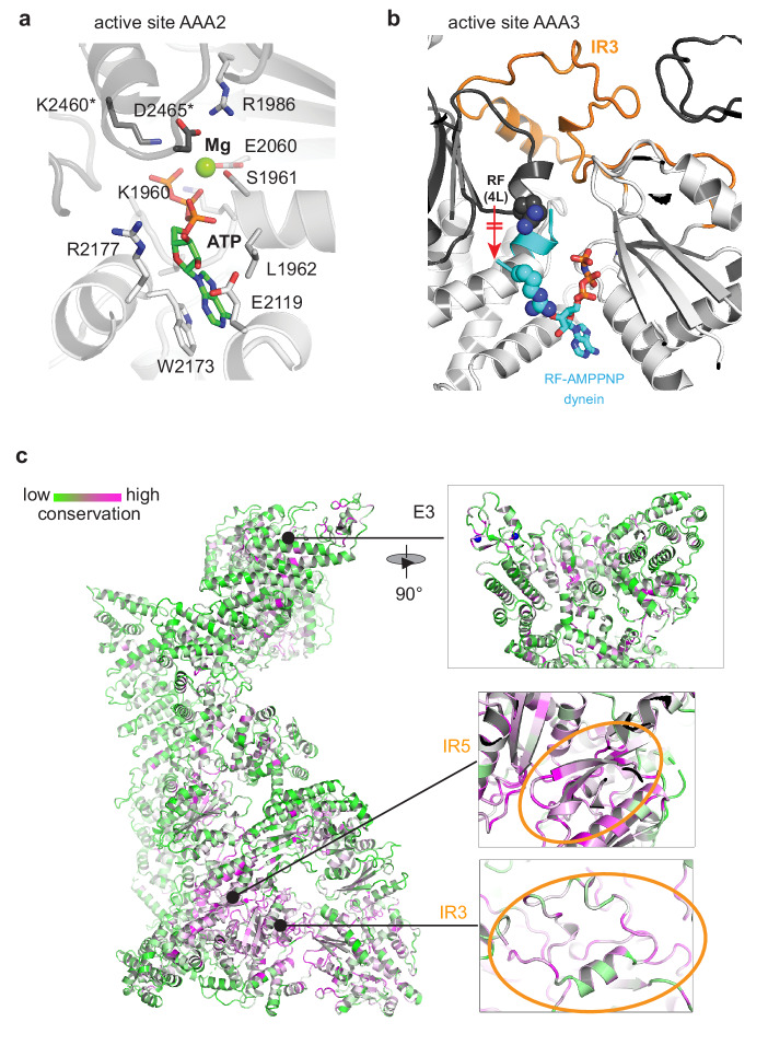
Figure 2—figure supplement 2. Conservation pattern of RNF213 mapped to the cryo-EM structure.

Figure 2—figure supplement 2.

(a) Atomic model of the AAA2 active site, tightly engaging ATP/Mg. Residues of the adjacent AAA rigid body are marked (*). (b) IR3 stabilizes the inactive state of AAA3 by trapping the arginine finger (RF) in a remote position from the AAA3 active site. For comparison, the aligned RF motif of the dynein AMPPNP structure (PDB ID 4w8f) is shown in cyan together with the bound nucleotide. (c) Conservation pattern of the full-length protein colored according to the degree of sequence conservation from low (green) to high (magenta). This plot illustrates that the core of the AAA ring represents the most highly conserved portion of RNF213. The zoomed-in inserts illustrate the conservation of the central E3 part, where only few functional motifs are conserved including the Zn-coordinating residues of the RING (top), the conservation of the IR5 motif (orange circle) and the conservation of the IR3 motif (orange circle). Notably, for IR3, specifically those residues that extend to the protein interior are conserved, whereas the conservation of surface-exposed residues varies.
Figure 2—figure supplement 3. Structural comparison of RNF213 and Rea1.

Figure 2—figure supplement 3.

The picture compares the overall organization of the two AAA6 proteins. From the aligned AAA rings, large C-terminal extensions project into the periphery. In case of RNF213, the tilted AAA6 domain guides the C-terminal E3 extension into a different direction aligning it with the N-arm, which is absent in Rea1.