Fig. 8.
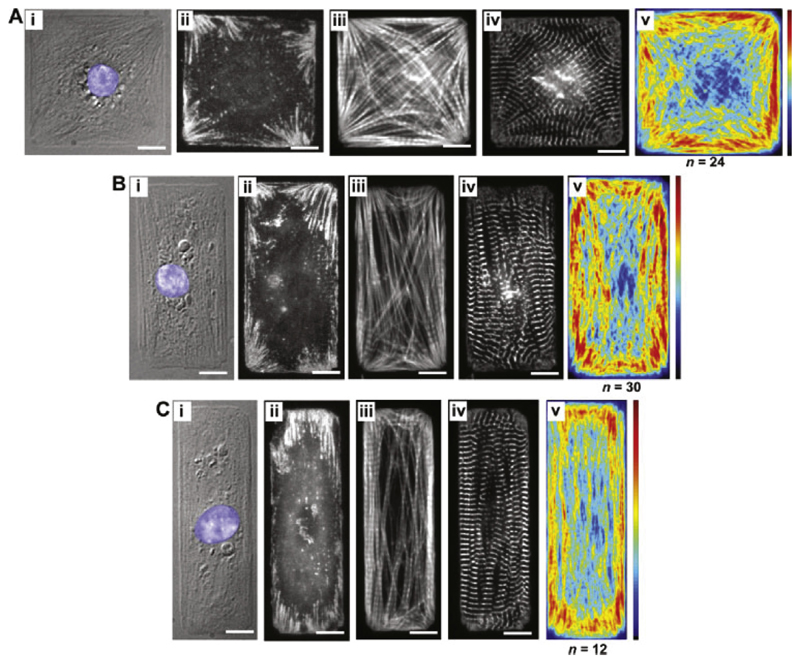
Bray et al. [135] printed ECM islands and placed myocytes on them to study the influence of the ECM on intracellular constituent alignment. The three cellular aspects are (A): 1:1, (B): 2:1 and (C): 3:1. (i) depicts a DIC image, (ii)–(iv) immunofluorescent stains for vinculin (revealing focal adhesions), F-actin (staining I-bands) and sarcomeric α-actin (revealing Z-bands). The average distribution of F-actin is shown in (v). Reproduced with permission from [135].

