FIGURE 2.
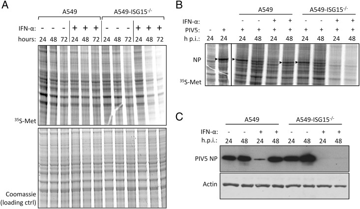
Analysis of cellular and viral protein synthesis in ISG15-deficient cells during an antiviral state. (A) Subconfluent A549 and A549-ISG15−/− (B8) cells were treated with 1000 IU/ml IFN-α or left untreated. At 24, 48, and 72 h, cells were pulsed for 1 h with L-35S-Met in Met-free media to metabolically label nascent proteins. Proteins were resolved by SDS-PAGE and stained with Coomassie to ensure equal loading. Labeled proteins were visualized by phosphorimager analysis. (B) A549 and A549-ISG15−/− (B8) cells were treated with 1000 IU/ml IFN-α for 8 h or left untreated and then infected with PIV5-W3 (MOI = 10). At 24 or 48 h p.i., cells pulsed and processed as in (A). Arrow heads denote 35S-Met–labeled PIV5 NP. Both experiments were performed independently at least twice. (C) PIV5-infected lysates from (B) were immunoblotted, and the accumulations of PIV5 NP and β-actin were detected with specific Abs and HRP-conjugated secondary Abs.

