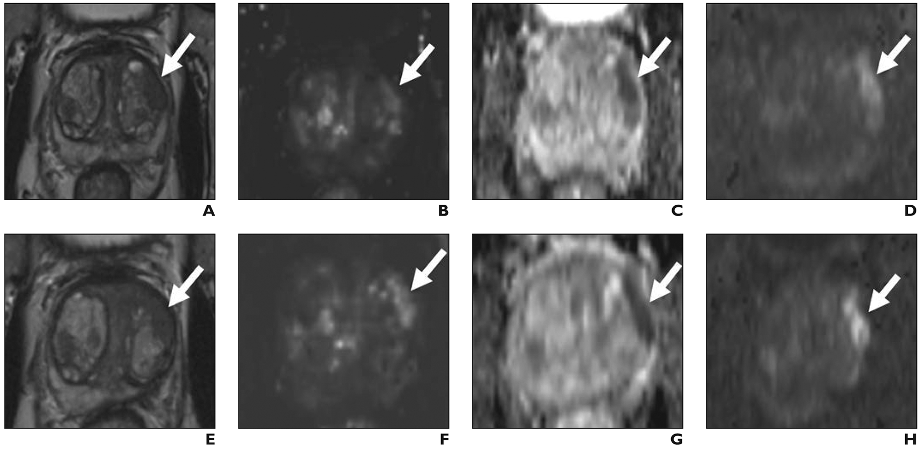
Fig. 1— 67-year-old man with prostate-specific antigen level of 8.4 ng/mL.

A–D, Before focal laser ablation, T2-weighted (A) and dynamic contrast-enhanced (B) images, apparent diffusion coefficient (ADC) map (C), and DW image (D) show Prostate Imaging Reporting and Data System version 2 5/5 lesion (arrow) in left anterior transition midgland corresponding to Gleason 3 + 4 prostate cancer at targeted biopsy.
E–H, Six months after focal laser ablation, T2-weighted (E) and dynamic contrast-enhanced (F) images, ADC map (G), and DW image (H) show persistent masslike low signal intensity, focal early enhancement, and markedly restricted diffusion (arrow). One radiologist scored T2-weighted, dynamic contrast-enhanced, and DW images 2, 2, 2; other radiologist scored them 1, 2, 2. Targeted biopsy 6 months after ablation revealed persistent Gleason 3 + 4 prostate cancer.
