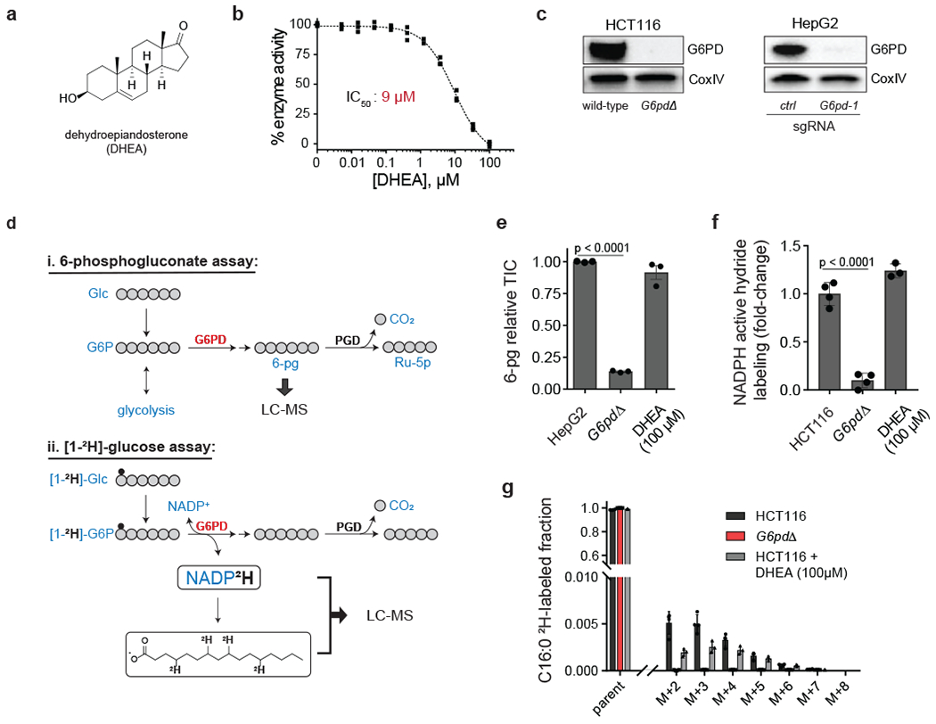
Figure 1. Cellular target engagement assays reveal lack of effective G6PD inhibition by DHEA.

a, Chemical structure of the steroid derivative dehydroepiandosterone (DHEA). b, In vitro activity of DHEA against recombinant human G6PD (mean ± SD, n = 3). c, Western blots of G6PD knockout cells generated using CRISPR-Cas9 (HCT116 knockout is clonal; HepG2 is batch; “ctrl” represents an intergenic control). See Supplementary Figure 16 for uncropped gels. Representative results of 2 independent experiments. d, Assays for G6PD cellular activity: (i) 6-phosphogluconate (6-pg) levels in HepG2 cells, (ii) deuterium (2H, small black circle) incorporation into NADPH (active hydride) and free palmitic acid from [1-2H]-glucose in HCT116 cells. e, f, g, DHEA (100 μM, 2 h) does not phenocopy G6PD knockout (TIC, total ion count by LC-MS) (mean ± SD, n = 3). p value calculated using a two-tailed unpaired Student’s t-test.
