
Figure 1.
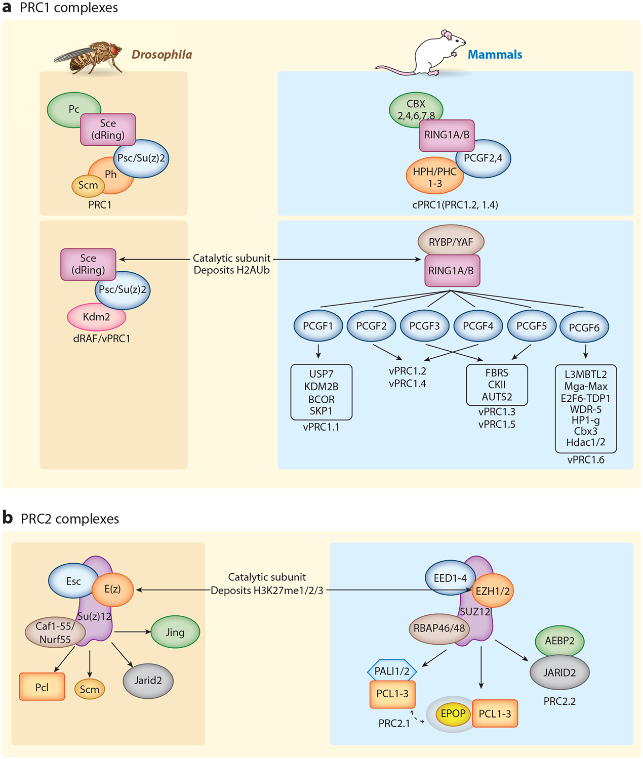
PcG complexes in Drosophila and mammals. PcG proteins are classified into two major complexes: (a) PRC1 and (b) PRC2. Homologous core complex subunits are color coded between Drosophila (left, brown) and mammals (right, blue), and their common catalytic subunits are indicated [dRing or RING1A/B in all PRC1 complexes and E(z) or EZH1/2 in PRC2]. The core complexes are diversified by interactions with accessory proteins, especially in mammals. Accessory subunits can be mutually exclusive as in mammalian PRC2.1 and PRC2.2. Similarly, mammalian PRC1 is divided into cPRC1 (canonical PRC1) and vPRC1 or ncPRC1 (variant or noncanonical PRC1), as initially defined by Gao et al. (38). Analogous in-depth studies of Drosophila vPRC1/ncPRC1 have not been reported. Protein–protein contacts presented here are not meant to be accurate. In many cases, they are not known in detail, although substantial progress has occurred recently (55–60, 81). Likewise, depicted complexes are meant to represent a general view, but the existence of additional configurations or cell type– and tissue type–divergent versions is also likely. Abbreviations: PcG, Polycomb Group; PRC1 and PRC2, Polycomb Repressive Complexes.