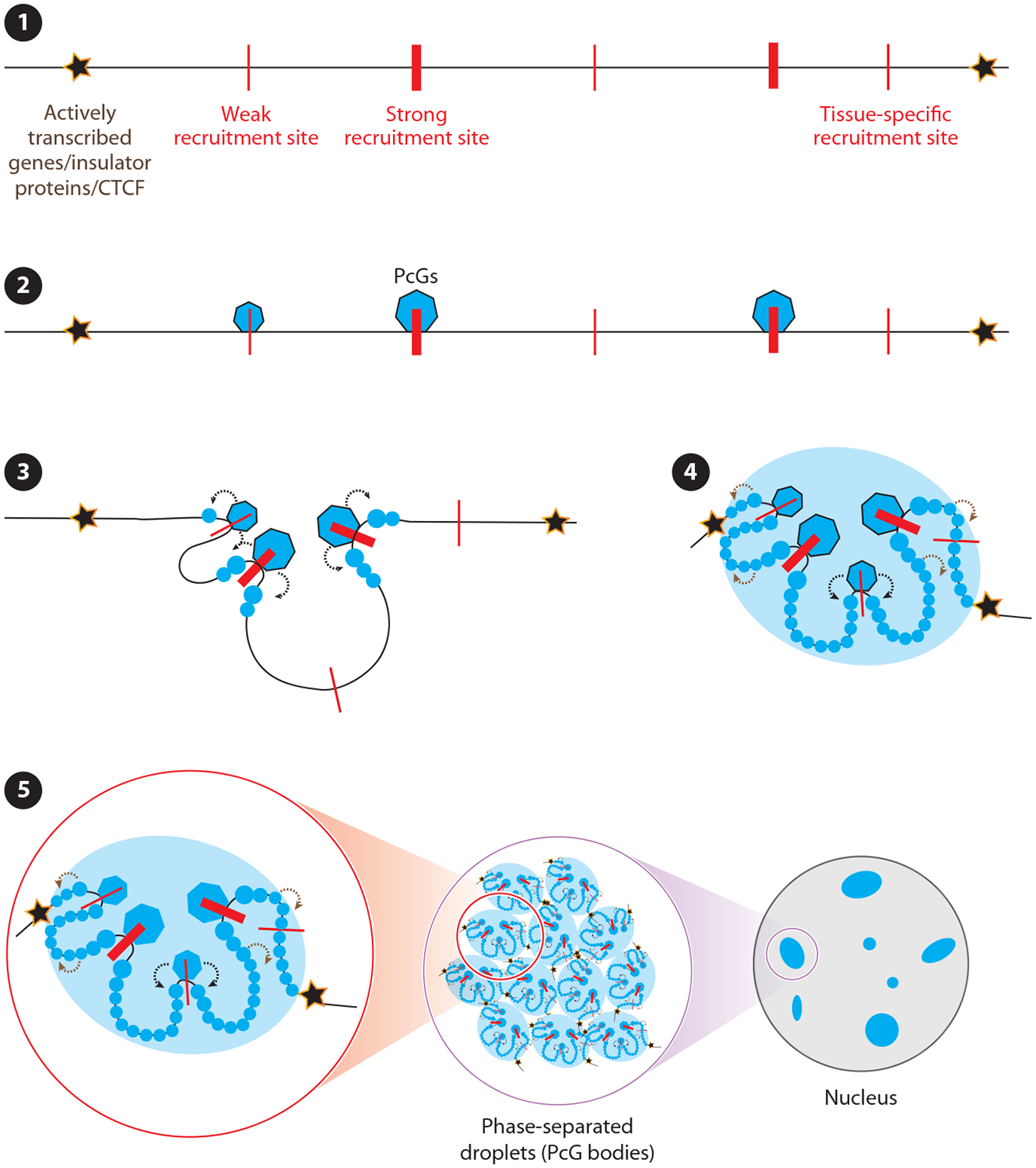
Figure 3.

Model for Polycomb Group (PcG) domain establishment. (❶) PcG domain boundaries are defined in Drosophila by either actively transcribed genes or insulator proteins and in mammals by CTCF proteins, and these boundaries may form similarly in flies and in mammals (3). (❷) In both organisms, PcG proteins appear to first engage with recruitment sites (red lines) within PcG target domains. (❸) The recruitment sites can be either strong or weak and either cell type–, tissue type–, or developmental stage–specific. After the initial recruitment, two parallel events happen: (❹) PcG complexes modify flanking histone tails (modified histones are shown with blue spheres) and interactions between the PcG proteins bound to the recruitment sites drive changes in 3D structure of the domain. The initial histone modifications and changes in 3D architecture of the domain drive further recruitment of PcG complexes and cause modification of the rest of the histones to establish the PcG domain. (➎) Finally, the PcG domains form phase-separated droplets either individually or by fusing with each other. These phase-separated droplets are also known as PcG bodies.
