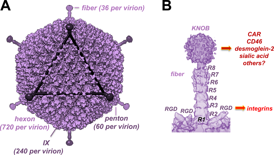
Figure 1. Cryo-Electron Microscopic Structures of Ad26.

A) Full virion structure B) Fiber and penton base. R indicated fiber shaft repeats. RGD indicates arginine-glycine-aspartic acid integrin binding motifs in the penton base. Knob indicates the receptor binding portion of the Ad26 fiber trimer. Receptors bound by these capsomers are shown on the right. Adapted from [33].
