. 2020 Feb 27;23(3):320–325. doi: 10.4048/jbc.2020.23.e18

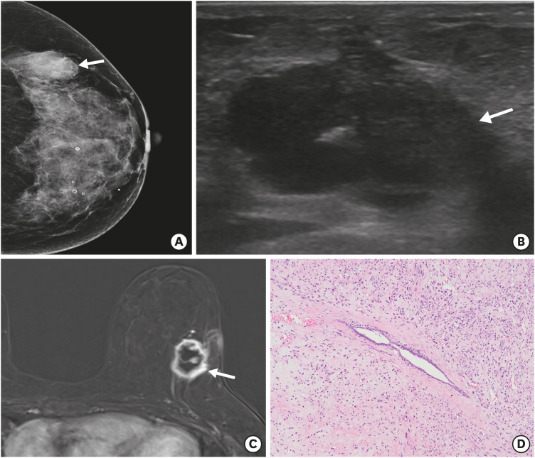
Figure 1. Malignant phyllodes tumor of the left breast at the time of initial diagnosis. (A) Craniocaudal view of the mammogram shows an indistinct high-density mass (arrow) in the left outer breast. (B) Ultrasound image shows an irregular hypoechoic mass (arrow) in the upper outer quadrant of the left breast. (C) Axial contrast-enhanced magnetic resonance image shows an irregular rim-enhancing mass (arrow) in the upper outer quadrant of the left breast. (D) Microscopically, the tumor shows a leaf-like pattern of cystic or cleft-like spaces lined with epithelium and abundant stromal cellularity (hematoxylin and eosin stain, ×100).

Figure 1