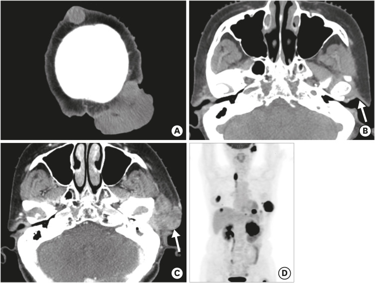
Figure 3. Widespread metastases of malignant phyllodes tumor. (A, B) A non-enhanced brain CT image for radiotherapy-planning shows increased size of the masses in the scalp. No abnormal mass lesion is observed in the left preauricular or parotid region (arrow in B). Eight months later, a rapid growing mass in the left preauricular area was noted. (C) Contrast-enhanced CT shows a heterogeneously enhancing mass (arrow in C) in the left parotid gland, with angioinvasion of the left external carotid artery and venous branches (not shown). (D) Maximum intensity projection reconstruction of a positron emission tomography image shows multifocal metastases in the left thoracic wall, left kidney, left lung and subphrenic space, left parotid gland, lymph nodes, and bones.
CT = computed tomography.

