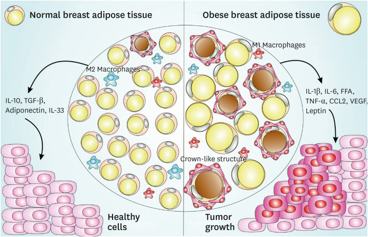
Figure 1. CLS-mediated signaling pathway in normal and obese adipose tissue and impact on breast cancer. Normal breast adipose tissue is characterized by the presence of smaller and less numerous white adipocytes, fewer crown-like structures, a prevalent M2 macrophages subset, with the secretion of anti-inflammatory cytokines such as IL-10, TGF-β, IL-33, and adiponectin. Obese breast adipose tissue is characterized by larger size and more abundant white adipocytes, with the secretion of pro-inflammatory cytokines such as IL-1β, IL-6, FFA, TNF-α, macrophages-chemoattractant CCL-2 chemokine, angiogenesis inducers such as VEGF, a prevalent M1 macrophages subset and an increased production of leptin, a negative feedback signal in the regulation of energy balance. This obese breast adipose tissue provides a favorable microenvironment to cancer establishment and progression.
CLS = crown-like structures; IL = interleukin; TGF = transforming growth factor; FFA = free fatty acid; TNF = tumor necrosis factor; CCL = chemokine (C-C motif) ligand; VEGF = vascular endothelial growth factor.

