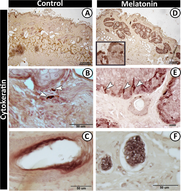
Figure 11.
Immunoreactivity of scrotal skin for cytokeratin protein-19 in control (A–C) and melatonin treated groups (D–F) showing; intense positive immunoreactivity of sebaceous glands in melatonin treated group (D) while those of control group (A) displayed negative immunoreaction. Strong immunoreactivity of the outer cells (arrowheads) of sebaceous glands (E), vascular elements (C) and nerve fibers (F) in melatonin treated group. Notice, the dendritic cells (arrowheads) exhibiting positive reaction for cytokeratin around the sebaceous glands in control group (B).

