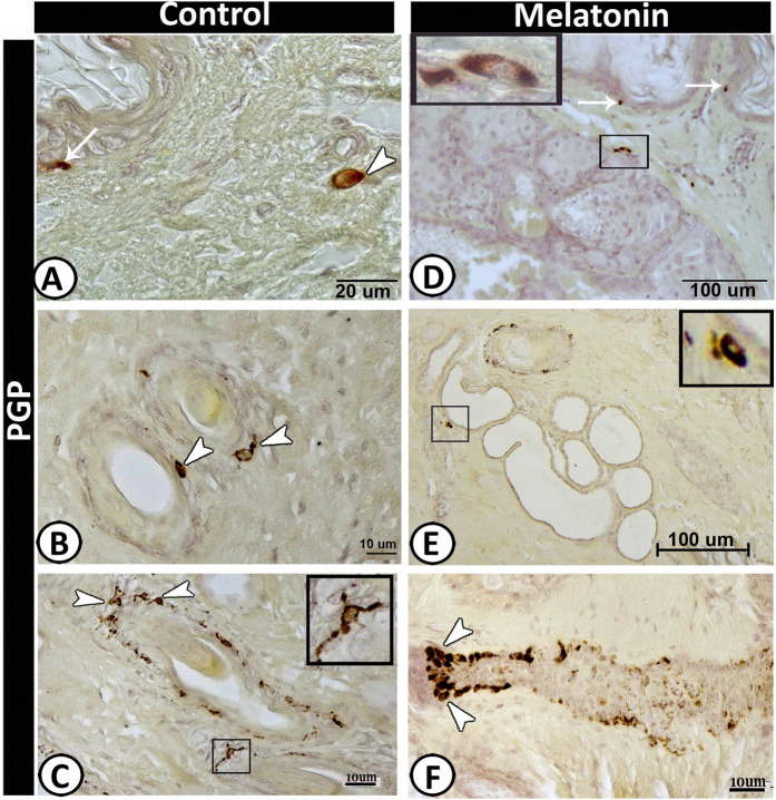
Figure 5.
PGP 9.5 immunoreactive cells in scrotal skin in control (A–C) and melatonin treated group (D–F). Langerhans cells (arrows) were observed in the basal layer of epidermis which were much frequent in melatonin group (D) than control (A). Dendritic cells (arrow) in the dermis in control group and around the hair follicle (B & F), the sebaceous gland (D) as well as sweat glands (selected area in E). Notice, TCs around the outer root sheath of the hair follicle (selected area in C).

