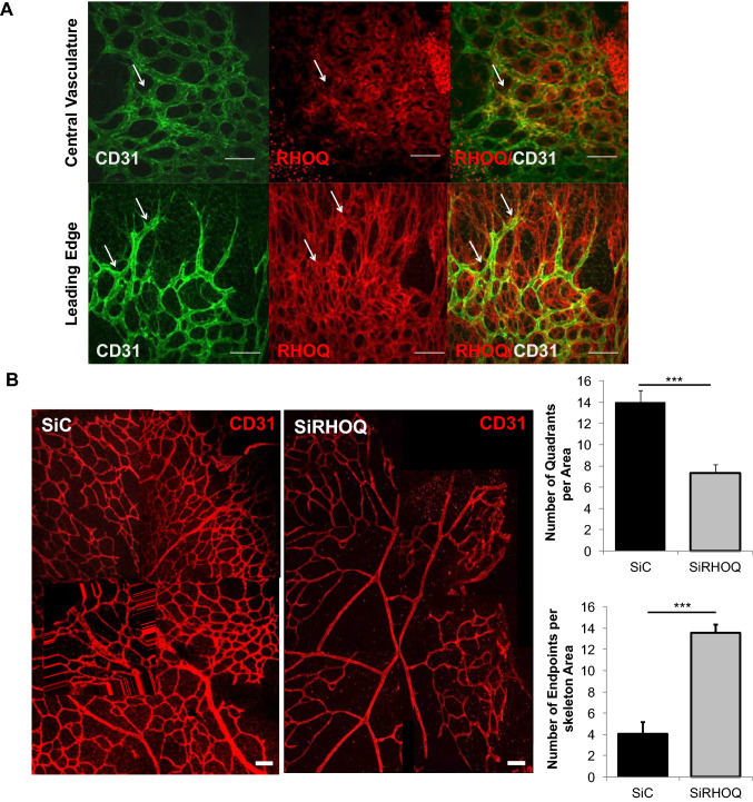
Fig. 4.
Loss of RHOQ disrupted retinal vasculature formation. a RHOQ (red) is expressed in endothelial cells (CD31, green), and other cell types, in the central vasculature and at the leading edge in retinas obtained from day 6 old pups. b Retinas were transfected following intraocular injection of 500 nM in 1 ul of SiControl (SiC) or SiRHOQ mouse duplexes using the in vivo GFP-labelled JETPEI delivery system on day 3 following birth of pups and eyes harvested on day 6 following birth and effects on vasculature assessed by immuno-fluorescence staining by analysis of quadrants formed at both central and leading edge vasculature and open endpoints of cells that failed to form a connection with quadrant in the central vasculature. (White arrow, example of co-localisation; Scale bar = 20 nm; data representative of n = 3 independent experiments)

