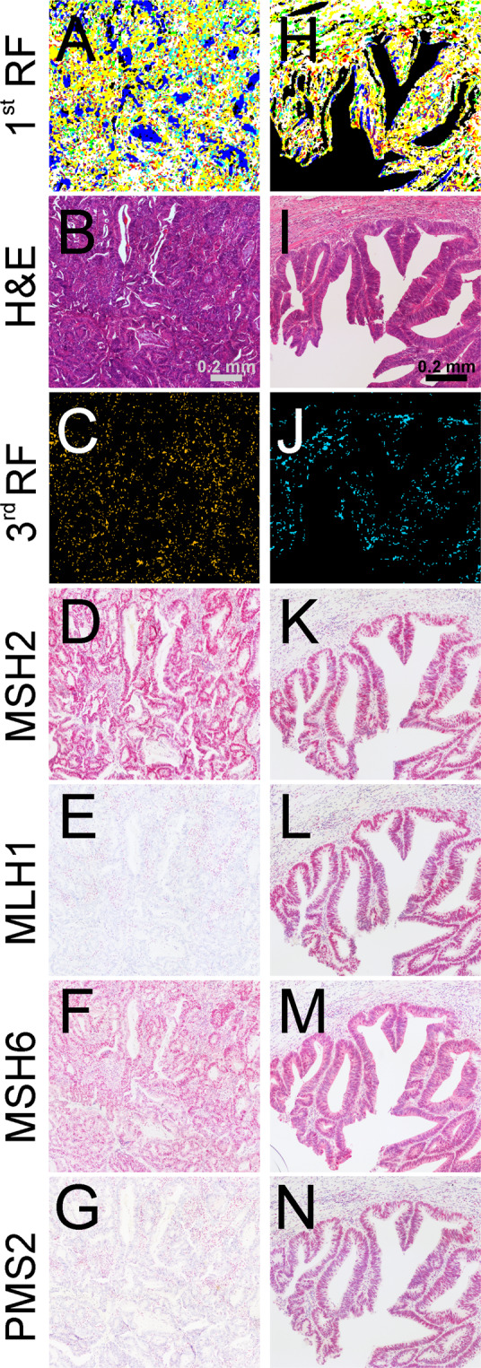
Figure 4.

Infrared (IR) imaging for a microsatellite instability-high (MSI-H) and stable (MSS) colorectal cancer (CRC) sample (light blue) compared to hematoxylin and eosin (H&E) staining and immunohistochemistry (IHC). IR imaging results of exemplary CRC tissue regions (A,C,H,J) are shown in comparison to H&E staining (B,I). Shown in (A,H) are muscle (white), infiltrating inflammatory cells (yellow), connective tissue (green), and pathologic regions (red) and in (C,J), microsatellite status is shown as follows: MSI-H in orange (C) and MSS in light blue (J). For more details on RFs, please refer to Fig. 1A. In D-G and K-N, the four IHC protein markers MSH2, MLH1, MSH6, and PMS2 are shown for adjacent tissue sections. For MSI-H, negative staining for MLH1 (E) and PMS2 (G) indicates the defect in these mismatch repair (MMR) proteins.
