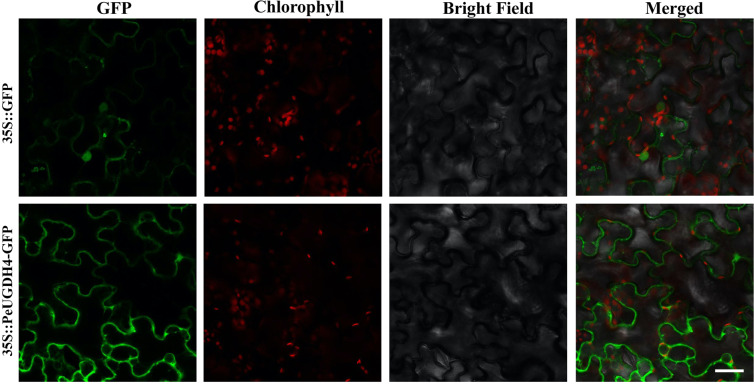
Figure 8.
Subcellular localization of PeUGDH4-GFP fusion protein in tobacco epidermic cells. Scale = 50 μm; the full-length ORF of PeUGDH4 was fused with GFP in pCAMBIA2300 plant expression vector drived by CaMV35S promoter (35S::PeUGDH4-GFP) and pCAMBIA2300-GFP vector (35S::GFP) was used as the positive control. Fluorescence signals are monitored by laser confocal microscopy, and the excitation light wavelengths are respectively: 488 nm for GFP and 663–738 nm for chlorophyll spontaneous fluorescence.

