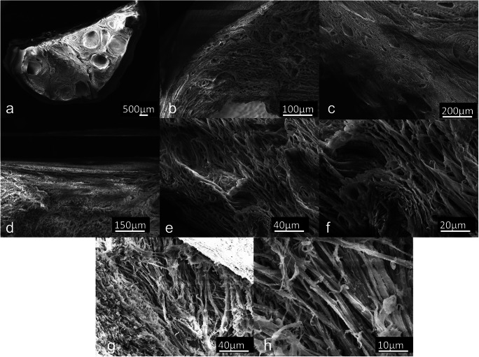
Fig. 2.
Scanning electron microphotographs of decellularized (DECELL) ovaries. a Decellularized hemiovary section. b, c Efficient cell removal, preservation of 3D microarchitecture, and ECM integrity are revealed after the decellularization process. Porous structures once populated by different cell types and complex fiber network are visible. d–h The ovarian surface epithelium and well-organized collagen fibers within pore walls are distinguishable

