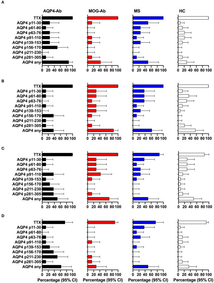
Figure 4.
CD4+ T-cell reactivity to AQP4 peptides in participants with AQP4-Ab (n = 8), MOG-Ab (n = 7), MS (n = 8) and HC (n = 14). (A) percentage of participants with positive CD4+ T-cell proliferation (CDI ≥ 3) after challenging with respective AQP4 peptides and the positive control TTX. Percentage of participants with positive secretion (SI ≥ 3) of GM-CSF (B), IFN-γ (C) and IL-6 (D) after challenging with respective AQP4 peptides and the positive control TTX. The 95% confidence intervals are indicated by the error bars. AQP4-Ab, aquaporin-4 antibody positive; CDI, cell division index; GM-CSF, granulocyte-macrophage-colony-stimulating factor; HC, healthy controls; IFN, interferon; IL, interleukin; MOG-Ab, myelin oligodendrocyte glycoprotein antibody positive; MS, multiple sclerosis; SI, stimulation index; TTX, tetanus toxoid.

