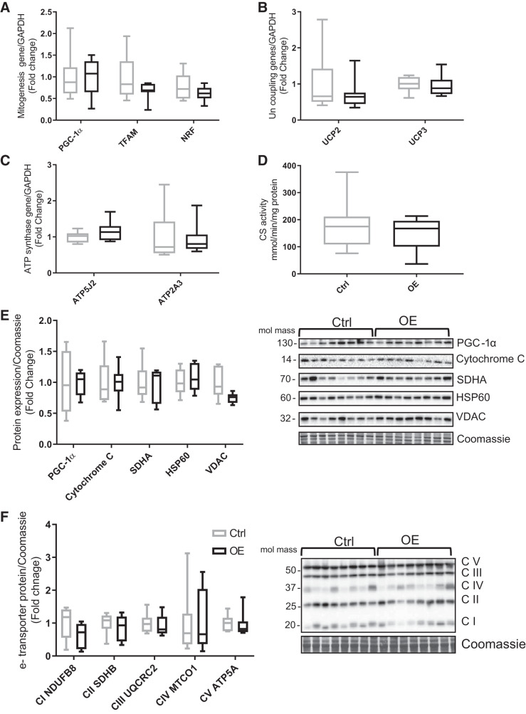
Fig. 4.
Markers of mitochondrial biogenesis and density in tibialis cranialis muscle. A: mitochondrial biogenesis regulatory genes: peroxisome proliferator-activated receptor-γ coactivator-1α (PGC-1α), mitochondrial transcription factor A (TFAM), and nuclear respiratory factors (NRF). B: gene expression of uncoupling proteins 2 and 3 (UCP2/3). C: ATP synthase genes ATP5J2 and ATP2A3. D: mitochondrial density represented by citrate synthase (CS) activity. E: protein levels of mitochondrial biogenesis markers: PGC-1α, cytochrome c, succinate dehydrogenase enzyme A (SDHA), heat shock protein 60 (HSP60), and voltage-dependent anion channel (VDAC). F: electron (e-) transporter complexes (C) of mitochondria: CI NADH dehydrogenase [ubiquinone] 1β subunit 8 (NDUFB8), CII succinate dehydrogenase subunit B (SDHB), CIII ubiquinone cytochrome c reductase complex (UQCRC2), CIV mitochondria encoded cytochrome c oxidase (MTCO1), and CV mitochondria membrane ATP synthase 5A (ATP5A). Values are means ± SD. Statistical analysis was via unpaired t test. No statistically significant differences were observed. Molecular mass is in kilodaltons. Ctrl, control group; OE, overexpressed group.

