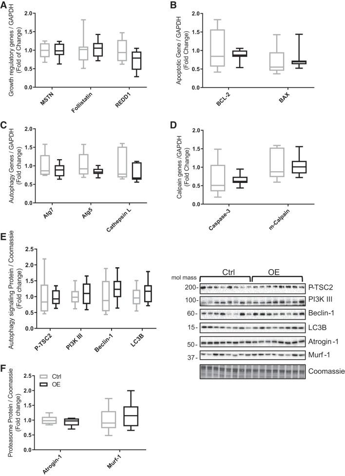
Fig. 6.
Gene expression levels in tibialis cranialis muscle of the following. A: negative growth regulatory factors: myostatin (MSTN), follistatin, and regulated in development and DNA damage response 1 (REDD1). B: apoptotic factors: B-cell lymphoma 2 (BCL-2) apoptosis suppressor and BCL-2-associated X (BAX) proapoptotic factor. C: autophagy-related genes: Atg7, Atg5, and cathepsin L. D: calpain factors: cysteine-aspartic acid protease (caspase-3) and m-calpain. E and F: protein expression of autophagy signaling pathway components: phosphorylated tuberin or tumor suppressor 2 (P-TSC2), phosphatidylinositol 3-kinase class III (PI3K III), beclin-1, autophagy marker light chain 3 (LC3B), atrogin-1, and muscle RING-finger protein-1 (Murf-1). Values are means ± SD. Statistical analysis was via unpaired t test. No statistically significant differences were observed. Molecular mass is in kilodaltons. Ctrl, control group; OE, overexpressed group.

