Fig. 1.
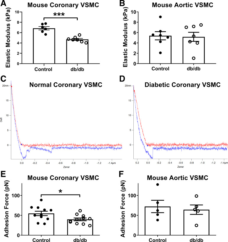
Diabetic mouse coronary vascular smooth muscle cells (VSMCs) are less stiff and less adhesive. A: elastic modulus, a measurement of cellular stiffness, was reduced in diabetic coronary VSMCs compared with normal. B: by comparison, there were no significant differences in elastic modulus between normal and diabetic aortic VSMCs. C: representative normal coronary VSMC force curve. D: representative diabetic coronary VSMC force curve. In C and D, red curve represents approach and blue curve represents retraction of the probe; therefore, x-axis is distance and y-axis is laser deflection. E: adhesive forces were reduced in diabetic coronary VSMCs compared with normal. F: by comparison, there were no significant differences in adhesive forces between normal and diabetic aortic VSMCs. n = 5–11 per group; *P < 0.05, ***P < 0.0001 vs. control. Statistical significance was assessed by unpaired Student’s t test.

